PAK1 (基因名), Serine/threonine-protein kinase PAK 1 (蛋白名), pak1_bovin.
产品名称:
Bovine PAK1/ Serine/threonine-protein kinase PAK 1 ELISA Kit
丝氨酸/苏氨酸蛋白激酶1
货号:
E13344b
商标:
EIAab®
监管等级:
别名:
Alpha-PAK, p21-activated kinase 1, p65-PAK, PAK-1
检测方法:
ELISA
特异性:
Natural and recombinant bovine Serine/threonine-protein kinase PAK 1
样品类型:
Serum, plasma, tissue homogenates, cell culture supernates and other biological fluids
样品数据:
登录.
研究领域:
Cancer
通用注释
亚单元:
Homodimer in its autoinhibited state. Active as monomer. Component of cytoplasmic complexes, which also contains PXN, ARHGEF6 and GIT1. Interacts with NISCH (By similarity). Interacts with DVL1; mediates the formation of a DVL1, MUSK and PAK1 ternary complex involved in AChR clustering (By similarity). Binds to the caspase-cleaved p110 isoform of CDC2L1 and CDC2L2, p110C, but not the full-length proteins. Interacts with ARHGEF7. Interacts tightly with GTP-bound but not GDP-bound CDC42/P21 and RAC1. Probably found in a ternary complex composed of DSCAM, PAK1 and RAC1. Interacts with DSCAM (via cytoplasmic domain); the interaction is direct and enhanced in presence of RAC1. Interacts with SCRIB. Interacts with PDPK1. Interacts (via kinase domain) with RAF1. Interacts with NCK1 and NCK2. Interacts with TBCB. Interacts with CRIPAK. Interacts with BRSK2. Interacts with SNAI1. Interacts with CIB1 (via N-terminal region); the interaction is direct, promotes PAK1 activity and occurs in a calcium-dependent manner.
功能:
Protein kinase involved in intracellular signaling pathways downstream of integrins and receptor-type kinases that plays an important role in cytoskeleton dynamics, in cell adhesion, migration, proliferation, apoptosis, mitosis, and in vesicle-mediated transport processes. Can directly phosphorylate BAD and protects cells against apoptosis. Activated by interaction with CDC42 and RAC1. Functions as GTPase effector that links the Rho-related GTPases CDC42 and RAC1 to the JNK MAP kinase pathway. Phosphorylates and activates MAP2K1, and thereby mediates activation of downstream MAP kinases. Involved in the reorganization of the actin cytoskeleton, actin stress fibers and of focal adhesion complexes. Phosphorylates the tubulin chaperone TBCB and thereby plays a role in the regulation of microtubule biogenesis and organization of the tubulin cytoskeleton. Plays a role in the regulation of insulin secretion in response to elevated glucose levels. Part of a ternary complex that contains PAK1, DVL1 and MUSK that is important for MUSK-dependent regulation of AChR clustering during the formation of the neuromuscular junction (NMJ). Activity is inhibited in cells undergoing apoptosis, potentially due to binding of CDC2L1 and CDC2L2. Phosphorylates MYL9/MLC2. Phosphorylates RAF1 at 'Ser-338' and 'Ser-339' resulting in: activation of RAF1, stimulation of RAF1 translocation to mitochondria, phosphorylation of BAD by RAF1, and RAF1 binding to BCL2. Phosphorylates SNAI1 at 'Ser-246' promoting its transcriptional repressor activity by increasing its accumulation in the nucleus. In podocytes, promotes NR3C2 nuclear localization. Required for atypical chemokine receptor ACKR2-induced phosphorylation of LIMK1 and cofilin (CFL1) and for the up-regulation of ACKR2 from endosomal compartment to cell membrane, increasing its efficiency in chemokine uptake and degradation. In synapses, seems to mediate the regulation of F-actin cluster formation performed by SHANK3, maybe through CFL1 phosphorylation and inactivation. Plays a role in RUFY3-mediated facilitating gastric cancer cells migration and invasion.
亚细胞位置:
Cytoplasm
Cell junction
Focal adhesion
Cell membrane
Cell projection
Ruffle membrane
Cell projection
Invadopodium
Recruited to the cell membrane by interaction with CDC42 and RAC1. Recruited to focal adhesions upon activation. Colocalized with CIB1 within membrane ruffles during cell spreading upon readhesion to fibronectin. Colocalizes with RUFY3, F-actin and other core migration components in invadopodia at the cell periphery (By similarity).
数据库链接
UniGene:
SMR:
STRING:
KEGG:
Pfam:
Uniprot:
该产品尚未在任何出版物中被引用。
[1].
牛丝氨酸/苏氨酸蛋白激酶1(PAK1)ELISA试剂盒可以做多少个样本?
牛丝氨酸/苏氨酸蛋白激酶1(PAK1)ELISA试剂盒分为2种规格,96孔和48孔。96孔的试剂盒,标曲和样本都做复孔的话,可以检测40个样本。96孔的试剂盒,标曲和样本都不做复孔的话,可以检测88个样本。
[2].
牛丝氨酸/苏氨酸蛋白激酶1(PAK1)ELISA试剂盒使用视频?
牛丝氨酸/苏氨酸蛋白激酶1(PAK1)ELISA试剂盒实验操作视频在以下网址中,对每一步的实验步骤都做了演示,方便实验员能更好地理解ELISA实验的过程。
https://www.eiaab.com.cn/lesson-tech/805.html
https://www.eiaab.com.cn/lesson-tech/805.html
[3].
牛丝氨酸/苏氨酸蛋白激酶1(PAK1)ELISA试剂盒是放在-20℃冰箱保存吗?
EIAab的牛丝氨酸/苏氨酸蛋白激酶1(PAK1)ELISA试剂盒,洗涤液、底物、终止液保存于4℃,其余试剂-20℃冰箱保存。
[4].
牛丝氨酸/苏氨酸蛋白激酶1(PAK1)ELISA试剂盒原理?
双抗体夹心法:用纯化的抗体包被微孔板,制成固相抗体,往包被有固相抗体的微孔中依次加入标准品或受检样本、生物素化抗体、HRP标记的亲和素,经过彻底洗涤后用底物TMB显色。用酶标仪在450nm波长下测定吸光度(OD值),计算样本浓度。
竞争法:用纯化的抗体包被微孔板,制成固相抗体,往包被有固相抗体的微孔中依次加入标准品或受检样本和生物素标记的目标分析物,受检标本中抗原与生物素标记抗原竞争结合有限的抗体。再加入HRP标记的亲和素,经过彻底洗涤后用底物TMB显色。用酶标仪在450nm波长下测定吸光度(OD值),计算样本浓度。
竞争法:用纯化的抗体包被微孔板,制成固相抗体,往包被有固相抗体的微孔中依次加入标准品或受检样本和生物素标记的目标分析物,受检标本中抗原与生物素标记抗原竞争结合有限的抗体。再加入HRP标记的亲和素,经过彻底洗涤后用底物TMB显色。用酶标仪在450nm波长下测定吸光度(OD值),计算样本浓度。
[5].
牛丝氨酸/苏氨酸蛋白激酶1(PAK1)ELISA试剂盒中需要使用的样品量是多少?
夹心法100μL/孔,竞争法50μL/孔。如样本浓度过高时,应对样本进行稀释,以使稀释后的样本符合试剂盒的检测范围,计算时再乘以相应的稀释倍数。
[6].
如何分析牛丝氨酸/苏氨酸蛋白激酶1(PAK1)ELISA试剂盒数据?
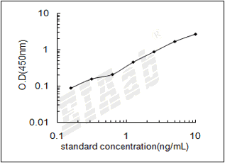
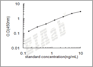
建议标准曲线,并计算样本浓度。对于elisa的曲线拟合,一般建议采用4参数曲线拟合,4参数曲线拟合通常更适合免疫分析。推荐使用专业软件进行曲线拟合,例如curve expert 1.3。根据样本的OD值由标曲查出相应的浓度,再乘以稀释倍数;或用标准物的浓度与OD值计算出标曲的回归方程式,将样本的OD值代入方程式,计算出样本浓度,再乘以稀释倍数,即为样本的实际浓度。以下链接是curve expert 1.3软件拟合曲线的方法。
https://www.eiaab.com.cn/news/502/
https://www.eiaab.com.cn/news/502/
[7].
牛丝氨酸/苏氨酸蛋白激酶1(PAK1)ELISA试剂盒中是否包含人和动物的副产物,是否包含感染的或者传染性原料如HIV等?
除了抗体和稀释液中的BSA,不含其它人和动物的副产物,也不含感染材料。
[8].
收集牛丝氨酸/苏氨酸蛋白激酶1(PAK1)ELISA试剂盒血浆样本,用什么作为抗凝剂?
一般建议用EDTA和肝素作为抗凝剂。
[9].
牛丝氨酸/苏氨酸蛋白激酶1(PAK1)ELISA试剂盒酶标板可以拆成几部分?拆的时候是否需要避光,无菌?
牛丝氨酸/苏氨酸蛋白激酶1(PAK1)ELISA试剂盒酶标板是8×12孔条,可拆卸,板子可以拆成12条,注意避免孔污染,不需要避光和无菌。暂时不用的板子,放回原来装的袋子里,密封保存。
[10].
牛丝氨酸/苏氨酸蛋白激酶1(PAK1)ELISA试剂盒样本如何保存?
尽量检测新鲜样本。若无新鲜样本,则4℃保存1周,-20℃保存1个月,-80℃保存2个月。
反馈墙
评论数 : 0
所有用户
所有用户
默认排序
默认排序
最近
早期
目前还没有评论。


通知
规格
数量
单价 (¥)
小计 1 (¥)
小计 2:
¥

规格
数量
单价 (¥)


 验证序列:
验证序列:


 折扣:
折扣: